Our Diagnostics division is dedicated to distributing and forming strategic partnerships with leading companies to deliver cutting-edge diagnostic solutions at the forefront of healthcare. Through collaboration with trusted innovators, we ensure the availability of reliable tools that enable precise disease detection, effective monitoring, and enhanced patient outcomes
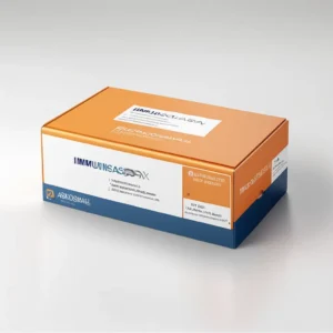
Purely human sera based – Mimics with human serum samples for results accuracy.
No extra additives added
compatible with all immunoassay analyzer ( Roche, Abbott, BC, Siemens, Mini Vidas, Snibe etc.)
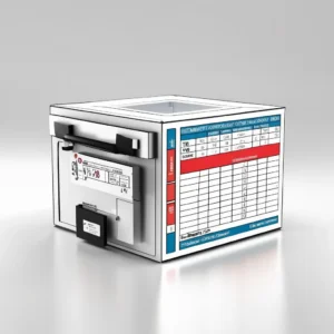
HEMATOLOGY CONTROLS ( 3 part & 5 part )
Monitoring of Analytical Process & detecting Errors
Ensuring Consistent Results:
Meeting Accreditation Requirements:
Multi-parameter controls for a wide range of hematological parameters, including erythrocytes, platelets, granulocytes, lymphocytes, monocytes, hematocrit, hemoglobin, and more.
Assayed Values:
Multiple Levels g., low, normal, high to assess the performance of the analyzer across a range of values.
Longer stability stability under proper storage conditions
Having compatibility with the specific hematology analyzers used in the laboratory.
Ready-to-Use control
Extended Differential Count: 5-part controls provide a more detailed breakdown of white blood cell types, including immature granulocytes.
Economical pack size to fit various segments of customers.
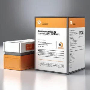
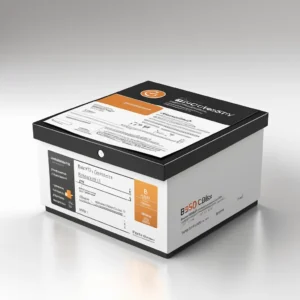
BIOCHEMISTRY CONTROLS
Assayed, lyophilized, and human serum-based, designed to monitor the precision of a wide range of chemistry assays.
Human Serum-Based mimicking the composition of patient samples.
Assayed: They have known values for specific analytes, allowing for verification of test accuracy.
Lyophilized: They are freeze-dried to ensure stability and long-term storage.
Internal Quality Control (IQC):
They are used as part of internal quality control (IQC) procedures, which involve using control materials and clinical samples during analysis to observe analytical errors.
Types of Controls – Normal Controls, Abnormal Controls , Routine Testing, Quality Control Systems: Improved Accuracy and Precision: By monitoring the performance of assays, controls help ensure that results are accurate and precise.
3-PART HEMATOLOGY ANALYZER
Electrical Impedance technology
Having aperture method used to determine cell volume and count.
Touchscreen display for ease of operation.
comprehensive database to store a large number of patient results
Having sample throughput of ~ 60 samples per hour
Features for quality control, such as Levey-Jennings charts and internal QC.
Compact Design for small to medium-sized clinics, laboratories, or physician offices.
Require only two reagents for operation.
Requires a small sample volume ~20 ul
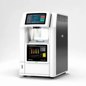
5-PART HEMATOLOGY ANALYZER
Differentiates white blood cells (WBCs) into five major subtypes (neutrophils, lymphocytes, monocytes, eosinophils, and basophils) using flow cytometry / Electrical Impedance/ Light Scattering/ Cytochemical Staining
5-part counters also analyze red blood cells (RBCs) and platelets (PLTs), providing a complete blood count (CBC) with a 5-part WBC differential.
Clinical Applications – Diagnosis and Monitoring, Infectious Disease, Hematological Malignancies
Having sample throughput of 60 – 80 samples per hour
Requires a small sample volume 25 – 55 ul
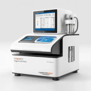
COAGULATION ANALYZER AND REAGENT
Test menu – Prothrombin Time (PT), Activated Partial Thromboplastin Time (APTT), Fibrinogen Levels, D-dimer , Factor Assays (for specific clotting factors) . Additional parameters Thrombin Time (TT) , Platelet Function Tests , Platelet Count
Precision and accuracy reduce the likelihood of human error.
Use of optical detection method.
Precise electronic pipettes for accurate reagents and sample dispensing.
Sample volume – 5 – 100 ul
Throughput – fit for various workload labs ranging for 100-400 test per hour.
Sample volume – ~ 500 ul and aspiration volume ~100 ul.
Sample type – EDTA or ESR vacuum tubes.
Optical technology or infrared light to analyse red blood cell sedimentations
Process – rouleaux formation, sedimentation, and packing.
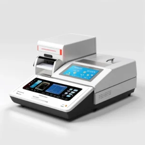
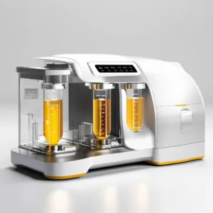
SEMI AUTOMATED BIOCHEMISTRY ANALYZER
Perform different types of chemical reactions, including kinetic, fixed-time, and endpoint assays.
Multipoint calibration and quality control to ensure accuracy and precision.
Ease of operation by Sample addition, holding, and colorimetric inhalation, as well as results recording and calculation including simple and intuitive user interface
Real-time Absorbance Monitoring
feature onboard reaction curve monitoring to visualize and analyze test results.
Multiple filters for different applications and wavelengths
Elimination of carry over contaminations between samples.
Sample volume – range of 1-100 ul.
Reagent volume – range of 1-400 ul
Throughput – ~ 150 test per hour
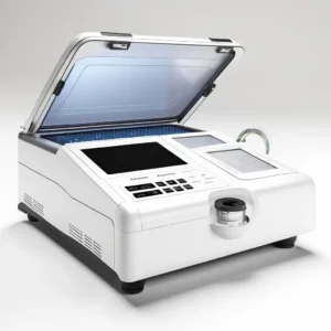
FULLY AUTOMATIC BIOCHEMISTRY ANALYZER
Test performed by spectrophotometry
Sample throughput ranging for 100-1200 tests per hour
Inbuilt ISE module optional
Automatic sample aspiration using single or double probe
Model available cuvette base or on board laundry
Manual intervention less or even no manual intervention,
More than 200 test parameters
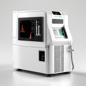
FULLY AUTOMATED IMMUNOASSAY MACHINE
High throughput, enhanced accuracy, and reduced manual intervention.
Handle a high volume of samples simultaneously, making them suitable for large laboratories or hospitals.
Random Access CLIA analyser
Built-in Quality Control to ensure the reliability of results.
Automated systems often optimize reagent usage, reducing waste and associated costs.
Throughput ranging from 200T/Hr.
Rreagents positions with more than ~20 nos..
Sample Positions with Automatic Barcode Scanning and Identification of ~50 nos.
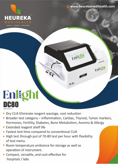
Broader test category – Inflammation, Cardiac, Thyroid, Tumor markers, Hormones, Fertility, Diabetes, Bone Metabolism, Anemia & Allergy
Extended reagent shelf life
Fastest test time compared to conventional CLIA
High test through put of 70-80 test per hour with flexibility of test menu.
Room temperature ambiance for storage as well as operation of instrument.
Compact, versatile, and cost-effective for hospitals / labs
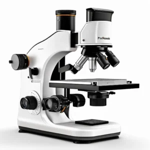
STUDENT MICROSCOPE
Simple, Compound & Molecular microscopes
Lightweight and compact for easy manoeuvre, storage and transport.
Durable construction for longer life
Portable and ease of mobility.
Objective lenses of 4x,10x&40x & eyepiece of 10x. Achromatic or semi plan.
Fixed or mechanical stages
Nosepiece: Revolving nosepiece for easy objective lens selection
Interpupillary Distance: Adjustable for binocular microscopes
Diopter Adjustment for individual eye correction
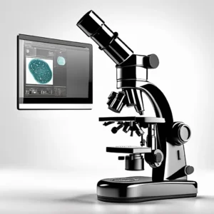
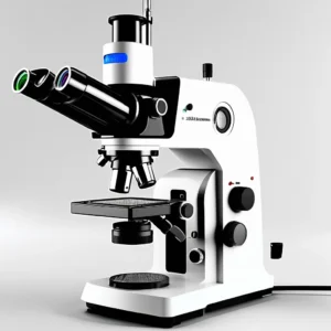
PATHOLOGIST MICROSCOPE
A bright-field compound microscope offer high resolution and magnification (40X-1000X), ergonomic features, and specific objective lenses (like 2x, 4x, 10x, 20x, 40x, and sometimes 60x) for detailed tissue analysis.
Magnification: Typically ranging from 40X to 1000X.
Oil immersion objectives for higher resolution.
Large field of view eyepieces (20 or 22 mm)
Ergonomics – Tiltable head for comfortable viewing.
Easy-to-use coarse and fine focus knobs.
Flip-in/flip-out condenser or diffusion slider for use with the 2x objective lens.
Mechanical stage for precise sample positioning.
Eyepiece Magnification: WF 10x/18mm.
Nosepiece: Triple, Upright.
FLUORESCENT MICROSCOPE
A fluorescent microscope enabling the study of cellular structures and processes.
Principle of Fluorescence: Excitation and Emission, Fluorophores, Wavelength Shift
It has key components i.e. Light Source, Excitation Filter, Dichroic Mirror, Emission Filter, Objective Lens
High Contrast and Specificity:
Fluorophores can be used to label specific molecules or structures, enabling researchers to visualize them in a sample.
Allows for the detection of multiple fluorophores in the same sample, enabling the study of complex biological systems.
Used to generate 3D images of samples.
Increase resolution allows for the visualization of smaller details.
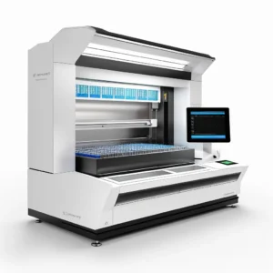
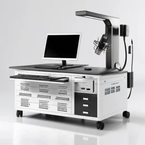
HISTOPATH + GROSSING STATION
HistoPath+ emerges as a modern digital solution, meticulously crafted to revolutionize the grossing process of surgical specimens and biopsies.
HistoPath+, operators embark on a journey of seamless digital documentation, capturing intricate details with unparalleled clarity.
HistoPath+ provides pathologists with access to high-resolution macro images, videos, and MP3 files, facilitating enhanced diagnostic reporting and collaboration.
Tissue sampling guidance
HistoPath+’s interactive interface and pre-loaded reporting features to facilitate integrated analysis and informed diagnoses.
Seamless navigation through detailed report data, formats, and comprehensive gross data for enhanced decision-making.
Automated measurements
Specimen tracking
Touchscreen interface
Safe formalin handling
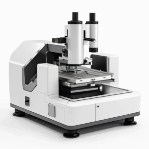
MICRODTOME
Blade Holder to hold securely the microtome blade, ensuring it remains in place during the cutting process.
The blade is clamped tightly to the holder, providing stability and allowing for precise control over the cutting angle and pressure.
Utilize low-profile blades, which minimize height or thickness, reducing vibrations and distortions during cutting for smoother, more accurate sections.
The specimen clamp firmly holds the tissue or material sample in place, preventing movement during sectioning.
Advance mechanism allows for precise control of the specimen’s movement towards the blade, enabling the cutting of thin, uniform sections.
Micrometer-driven specimen advance, enabling accurate control over section thickness.
It include safety features like blade guards and blade ejectors to enhance operator safety.
Electric cooling devices help to obtain uniform sections.
LCD Display with Feather Touch Keypad Slicing/Trimming mode
Able to provide section thickness range of 0.5 micron to 100 micron.
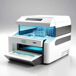
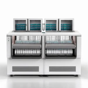
AUTOMATED TISSUE PROCESSOR
Feather touch key pad.
PID Controller controlled from main LCD screen.
Digital display for adjusting time sequence.
Carousel type tissue processor with 12 stations of 1.0/2.0 litre each; 10 reagent stations, 2 wax baths.
Fixation, Dehydration, Paraffin infiltration of histological tissue specimens with fixatives, alcohol, solvents and paraffin wax.
Vertical agitation or spiral or centrifugal with definite cycles.
Battery back-up to retain data.
MS Powder coated with anti-rusting chemical coating.
12 stage timing sequence can be programmed with each step of 1 minute to 999 minutes.
Vacuum pump is integrated in the instrument to minimize the contamination of reagent fumes in the lab environment.
Delay action of 1 hour to 99 hours.
2 electronically controlled wax bath 10oC to 80oC, accuracy of + 1oC.
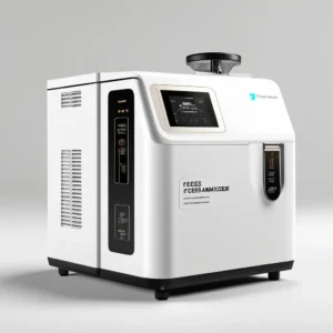
FECES ANALYZER
Feces analyzers, also known as stool analyzers, are instruments that perform various tests on stool samples to detect a range of health conditions, including infections, digestive issues, and cancers, by analyzing physical characteristics, microscopic elements, and chemical components.
Physical Examination -Analyzes the stool’s color, consistency, shape, and odor.
Microscopic Examination – Detects and identifies parasites, their eggs, and other microscopic elements.
Chemical Tests – to check substances like blood (occult blood), fat, mucus, and undigested food particles.
Bacterial and Viral Cultures – Detects the presence of bacteria, viruses, and fungi that cause infections.
Fecal Occult Blood Test (FOBT) – Detects hidden blood in the stool, which can be an indicator of colorectal cancer or other gastrointestinal conditions.
Stool pH test – measures the acidity or alkalinity of the stool, which can indicate certain digestive issues.
Automatic analyser for speed and efficiency & reduces turnaround time.
URINE ANALYZER
Fully automated urine analyzers offer features like high throughput, automated sample handling, and digital image analysis for efficient and accurate urinalysis, reducing manual labor and improving standardization.
High Throughput and Efficiency – upto ~ 240 samples / hour
Instrument performs features like automatic sample aspiration, reading, and reagent dispensing ensure consistent and reliable results.
Automated systems use algorithms to classify urine particles, ensuring consistent and objective results.
Digital Image Analysis by capturing images of urine sediment, allowing for detailed review and analysis by technicians.
Chemical and Physical Parameter Analysis
Analyzers can measure a wide range of chemical and physical parameters, such as pH, protein, glucose, ketones, and specific gravity.
Incorporate flow cytometry or automated microscopy to identify and count various urine particles, including red blood cells, white blood cells, and casts. & also differentiate between various types of urinary particles, such as different types of epithelial cells, crystals, and casts.